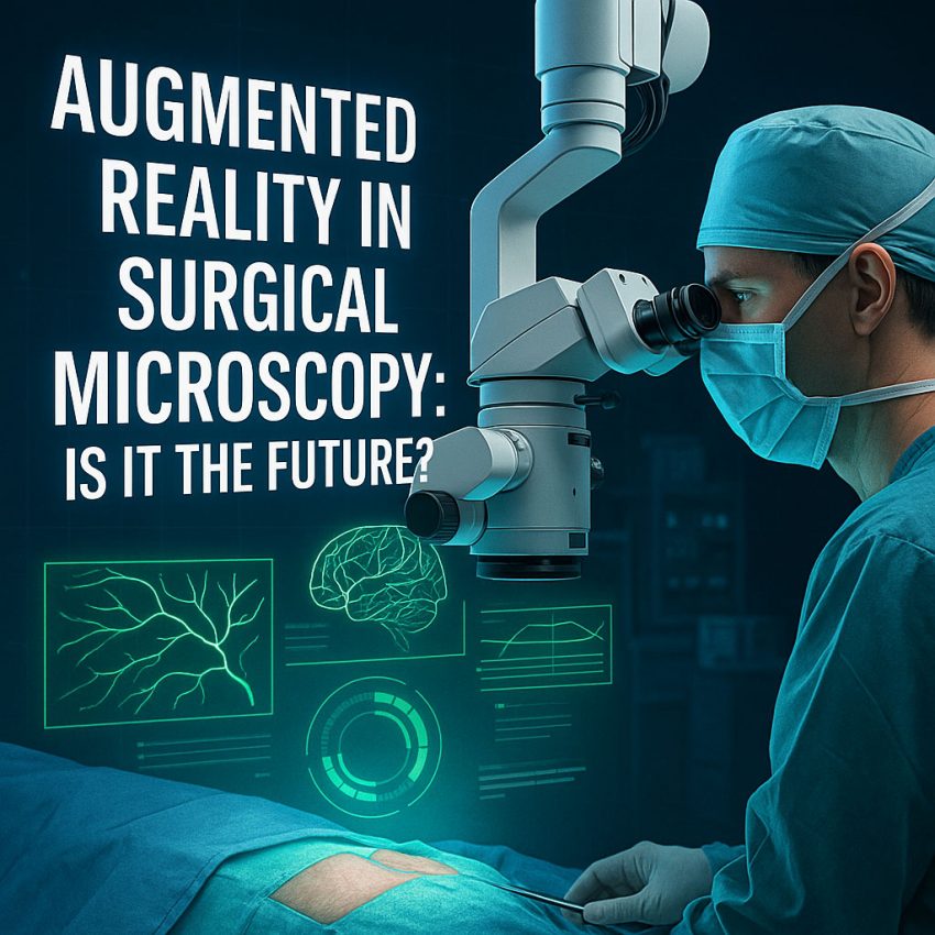
Surgical technology is evolving at a rapid pace, and among the most exciting advancements is the integration of Augmented Reality (AR) into surgical microscopy.
While surgical microscopes already provide magnified, high-resolution images of the operative field, the addition of AR overlays is changing the way surgeons visualize, plan, and execute complex procedures.
But is AR in surgical microscopy just another high-tech feature—or is it a glimpse into the future of precision surgery?
What Is Augmented Reality in Surgical Microscopy?
Augmented Reality (AR) refers to the real-time overlay of digital information—such as images, labels, or data—on the surgeon’s view of the physical world. In surgical microscopy, this means that critical data can be projected directly into the eyepiece or onto a display screen during surgery.
Examples of AR elements include:
Blood vessel maps
Tumor boundary outlines
Navigation paths
Depth cues
Anatomical labels
Perfusion indicators
These overlays are pulled from CT, MRI, or ultrasound data and aligned in real time with the live surgical view.
How Does It Work in Surgical Microscopes?
AR-enhanced surgical microscopes integrate multiple systems:
Imaging and Navigation – Preoperative imaging data is loaded into a navigation system.
Microscope Integration – The microscope receives real-time tracking of instrument location and patient anatomy.
Digital Overlay System – AR data is projected through the microscope’s optics or displayed on a monitor in sync with the surgical field.
User Controls – Surgeons can adjust the overlay opacity, layers, and focus through foot pedals or touch interfaces.
This fusion of real and digital visuals allows surgeons to operate with “X-ray vision,” seeing beyond what the naked eye can detect.
Benefits of AR in Surgical Microscopy
1. Enhanced Surgical Precision
AR provides clear anatomical references, reducing guesswork and helping surgeons avoid critical structures such as arteries, nerves, or tumors.
2. Real-Time Guidance
Live feedback supports better decision-making and reduces the likelihood of complications, especially in delicate brain, spinal, or ENT surgeries.
3. Minimally Invasive Efficiency
Surgeons can perform complex procedures through smaller incisions, guided by AR maps, reducing patient trauma and recovery time.
4. Educational and Collaborative Value
AR-enhanced views are displayed on monitors, making them invaluable for training, live-streaming, and team-based procedures.
5. Integration with Robotics and AI
AR can complement robotic surgery platforms, providing enhanced situational awareness and smart alerts powered by artificial intelligence.
Challenges and Limitations
1. Calibration and Accuracy
Misaligned overlays can pose risks. Precision tracking and registration are essential for safe outcomes.
2. System Complexity
Integration between navigation, imaging, and optical systems requires coordination and technical expertise.
3. Cost
AR-capable surgical microscopes and navigation systems represent a significant investment for hospitals.
4. Surgeon Adaptation
There’s a learning curve for surgeons unfamiliar with digital overlays or mixed-reality environments.
Current Applications in AR Surgical Microscopy
Neurosurgery: AR shows tumor margins, brain fiber tracts, and blood vessels during tumor resection.
Spine Surgery: Assists in pedicle screw placement and neural protection.
ENT & Skull Base Surgery: Navigates narrow anatomy with real-time spatial cues.
Plastic and Reconstructive Surgery: Visualizes vascular flow and flap design.
Ophthalmology: Supports precision incisions and intraocular navigation.
The Future of AR in Surgery
The integration of AI, AR, and 3D visualization is pushing the limits of surgical innovation. In the coming years, we can expect:
Voice-activated AR controls
Augmented overlays from intraoperative imaging (e.g., real-time ultrasound)
Wearable AR systems (headsets instead of microscopes)
Cloud-based AR surgical planning platforms
Some companies are already developing autostereoscopic displays and smart microscopes that adapt overlays based on the procedure’s progress.
Conclusion: Is AR the Future of Surgical Microscopy?
The answer is a resounding yes—with conditions. AR is not a gimmick; it’s a game-changer that enhances safety, precision, and education. However, widespread adoption will depend on cost-effectiveness, surgeon training, and seamless system integration.
As technology matures, Augmented Reality will likely become a standard feature of surgical visualization tools—ushering in a new era of intelligent, immersive, and intuitive surgery.
