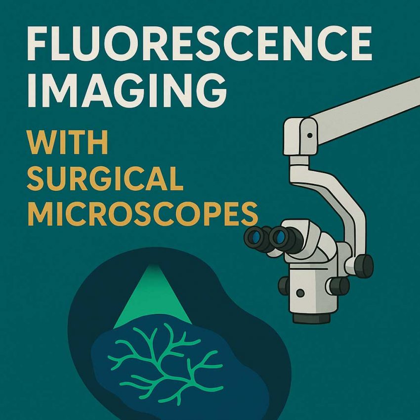
Discover how fluorescence imaging with surgical microscopes enhances surgical precision, improves visualization, and supports safer outcomes in modern medicine
Fluorescence imaging is rapidly transforming the way modern surgeries are performed. By enabling surgeons to visualize anatomical structures and physiological functions in real time, fluorescence imaging—when combined with advanced surgical microscopes—offers a level of precision that was once impossible with traditional techniques.
What is Fluorescence Imaging in Surgery?
Fluorescence imaging involves the use of fluorescent dyes or agents that emit light when exposed to a specific wavelength of light. When these agents are introduced into the patient’s body—typically through injection—they bind to specific tissues or circulate in the bloodstream. Under a specially equipped surgical microscope, these areas “glow” with a distinct color, allowing the surgeon to identify structures such as tumors, blood vessels, lymph nodes, and damaged tissues.
The most commonly used agent is indocyanine green (ICG), a dye that fluoresces under near-infrared (NIR) light. It’s safe, FDA-approved, and highly effective in numerous surgical applications.
How Surgical Microscopes Enable Fluorescence Visualization
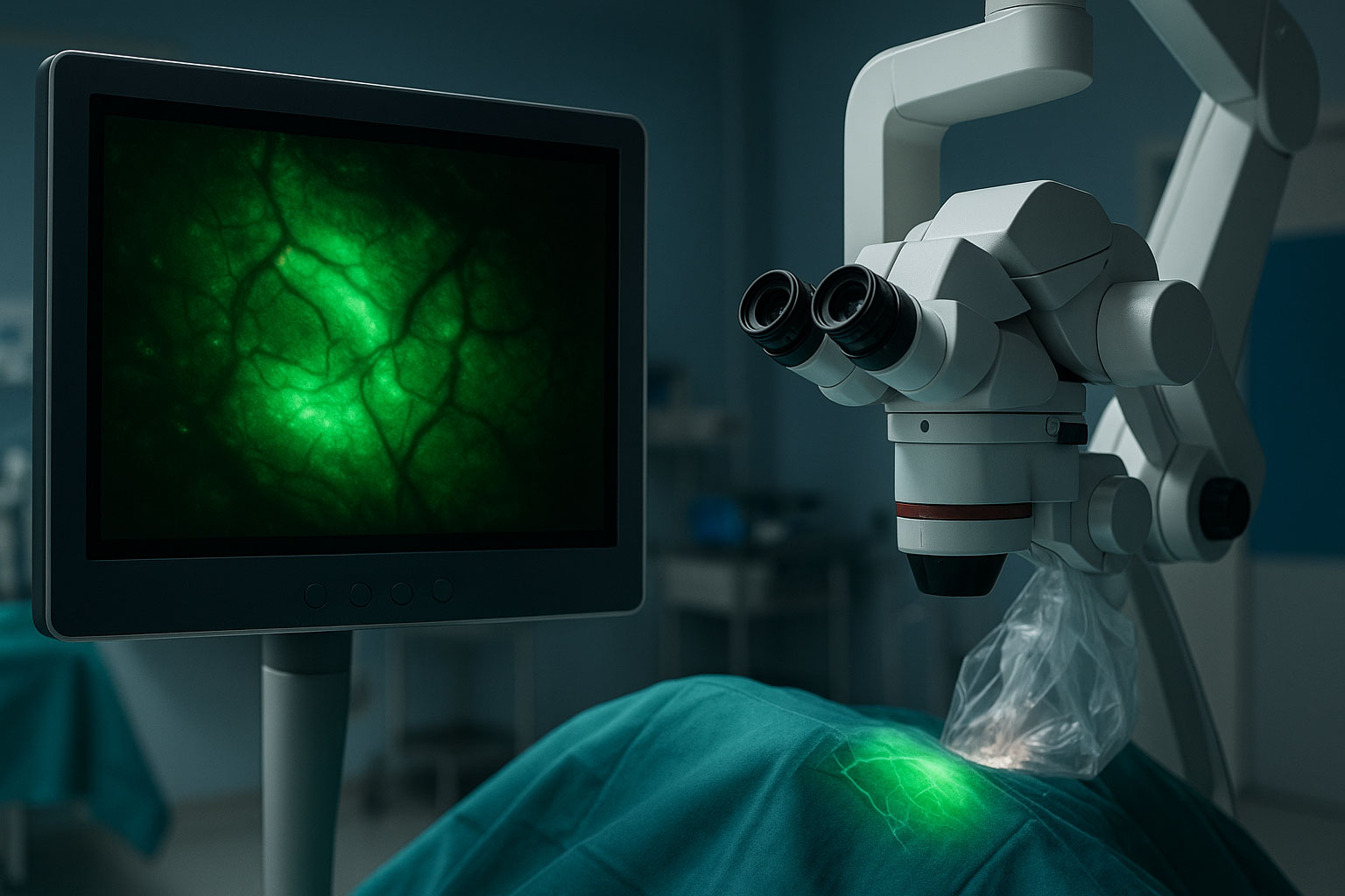
Modern surgical microscopes are equipped with specialized optics, cameras, and illumination systems that support fluorescence imaging. These microscopes allow the surgeon to toggle between standard white light and fluorescence modes at the press of a button—without interrupting the procedure. The result is a real-time, high-resolution view of fluorescent-marked tissues directly in the surgical field.
Some advanced models even overlay the fluorescent signal onto the white light image or display it side-by-side on digital monitors, giving the surgeon multiple views simultaneously.
Key Clinical Applications
1. Neurosurgery
In brain tumor surgery, dyes like 5-ALA or ICG highlight tumor cells, making it easier to distinguish between malignant tissue and healthy brain matter. This increases the likelihood of a complete resection while minimizing harm to surrounding functional areas.
2. Oncology
In cancer surgeries (such as breast, gastrointestinal, or gynecological procedures), fluorescence imaging helps surgeons visualize tumor boundaries, sentinel lymph nodes, and metastatic spread. It reduces the chances of leaving behind residual cancer cells.
3. Vascular and Cardiothoracic Surgery
ICG fluorescence is used to confirm vessel patency, detect blockages, and assess graft quality. This is especially useful in bypass surgeries or reconstructive vascular procedures.
4. Plastic and Reconstructive Surgery
Surgeons use fluorescence to evaluate the blood supply to skin flaps and grafts, helping ensure the tissue will survive after transplantation. This improves surgical outcomes and decreases the risk of necrosis.
5. Hepatobiliary and Gastrointestinal Surgery
Fluorescence imaging assists in identifying bile ducts and blood vessels during liver or gallbladder surgeries. It also helps in locating leaks or verifying the viability of anastomoses in gastrointestinal reconstructions.
Advantages of Fluorescence-Guided Microsurgery
- Greater Surgical Precision: It highlights vital structures, allowing for safer dissection and resection.
- Improved Decision-Making: Surgeons receive immediate visual feedback on tissue viability or tumor margins.
- Reduced Complications: By avoiding critical structures and confirming blood flow, the risk of postoperative issues decreases.
- Better Patient Outcomes: Complete tumor resections and successful flap integrations lead to shorter recovery times and better prognoses.
- Minimally Invasive Opportunities: With the help of real-time imaging, procedures can often be performed through smaller incisions, reducing trauma.
Limitations and Considerations
Despite its promise, fluorescence imaging has some challenges:
- Cost: Equipment and fluorescent agents can be expensive, potentially limiting widespread use in low-resource settings.
- Learning Curve: Interpreting fluorescence signals and integrating them into surgical decision-making requires training and experience.
- Depth Limitations: Fluorescence light cannot penetrate deeply into tissues, making it suitable mainly for surface or near-surface imaging.
- Timing and Dose Sensitivity: The effectiveness of fluorescence depends on the correct timing and dosing of the dye, which varies by procedure.
Emerging Trends and the Future of Fluorescence Microsurgery
Technological innovation continues to expand the potential of fluorescence imaging in surgery. Some trends to watch include:
- AI Integration: Artificial intelligence can enhance signal interpretation, automate structure detection, and support real-time decision-making.
- Multi-spectral Imaging: Future microscopes may use multiple fluorescent dyes simultaneously to target different structures or cell types.
- Augmented Reality (AR): Fluorescence signals could be projected onto AR displays, helping surgeons see “beneath” the surface of tissues.
- Wider Accessibility: As production scales up and costs decrease, more hospitals—especially in developing regions—may adopt these systems.
Conclusion
Fluorescence imaging with surgical microscopes is a milestone in the evolution of precision surgery. By enhancing visibility, guiding decisions, and supporting safer, more effective procedures, it is reshaping the surgical landscape across disciplines. As technology continues to evolve, this technique will likely become a gold standard in operating rooms around the world, offering benefits not just for surgeons—but more importantly, for the patients they serve.