What Is a Surgical Microscope? A Complete Beginner’s Guide to One of the Most Important Tools in Modern Surgery
In the world of modern medicine, precision can mean the difference between success and failure. Surgeons today rely on tools that allow them to operate with microscopic accuracy—and one of the most essential of those tools is the surgical microscope.
But what exactly is a surgical microscope? How does it work? What are its parts, and why is it so vital in surgery?
This guide will walk you through everything you need to know about surgical microscopes—from their basic function and structure to their real-world applications in medicine.
Definition: What Is a Surgical Microscope?
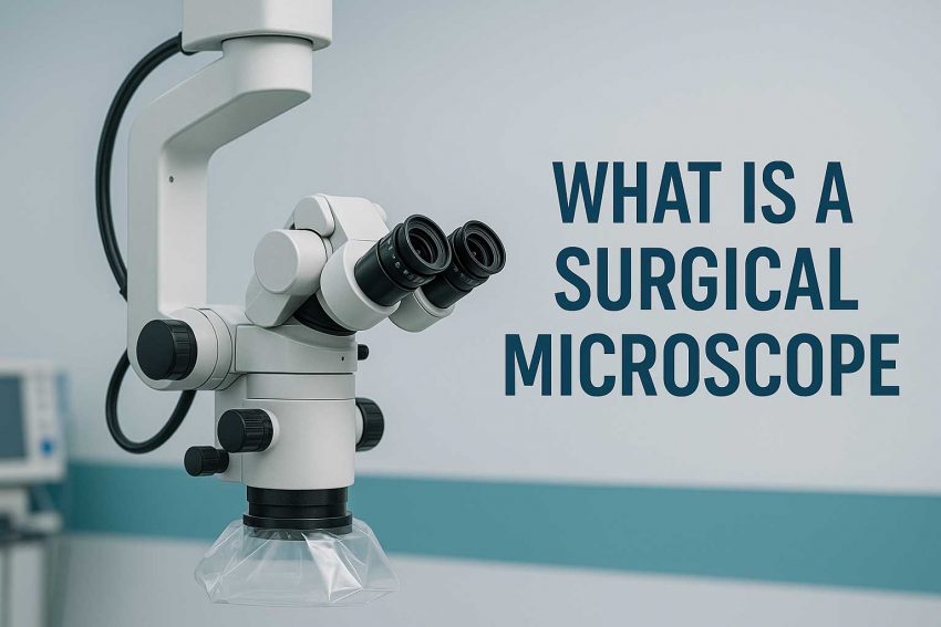
A surgical microscope is a high-precision optical instrument designed to provide magnified visualization of small structures during surgical procedures. Unlike simple magnifying lenses, surgical microscopes deliver:
Enhanced depth perception
Bright, focused illumination
Variable zoom
Hands-free operation
They are commonly used in surgeries that involve delicate tissues, narrow spaces, or minute structures, such as:
Neurosurgery
ENT (Ear, Nose, Throat)
Ophthalmology
Dental and endodontic surgery
Plastic and reconstructive surgery
Veterinary surgery
Key Components of a Surgical Microscope
Here are the primary parts that make up a modern surgical microscope:
| Component | Function |
| Binocular eyepieces | Allow the surgeon to view the surgical field with both eyes |
| Objective lens | Focuses light onto the surgical site and determines working distance |
| Zoom system | Provides variable magnification |
| Illumination system | Uses halogen, LED, or xenon light for shadow-free illumination |
| Stand/Base | Supports the microscope and allows adjustment during surgery |
| Foot controls | Lets surgeons adjust focus or zoom hands-free |
| Camera adapter | (Optional) For recording or streaming procedures for training or records |
Modern microscopes may also include digital imaging, 3D visualization, or robotic assistance.
How Does a Surgical Microscope Work?
Surgical microscopes use a combination of optical lenses and powerful light sources to create a magnified, highly detailed view of the area being operated on.
Here’s a simplified breakdown of how it works:
Light enters through the illumination system and is directed toward the surgical field.
Reflected light from the tissues passes through a series of lenses that magnify and clarify the image.
The surgeon views the image through the eyepieces, maintaining a comfortable posture and a high level of control.
Many surgical microscopes offer adjustable working distances, multiple zoom levels, and focus settings to adapt to the needs of different specialties and procedures.
Where Are Surgical Microscopes Used?
- Neurosurgery: Used for operations on the brain and spinal cord where the margin for error is zero. Precision and stability are critical.
- ENT Surgery: Essential in ear and sinus surgeries where small anatomy demands extreme magnification.
- Ophthalmology: Used in procedures like cataract removal and corneal surgery. The optics must be crystal-clear.
- Dental / Endodontics: Helps dentists and endodontists see inside root canals and perform intricate tooth surgeries.
- Veterinary Surgery: Used in advanced animal surgeries, especially in small animals where organs and tissues are tiny.
A Brief History of Surgical Microscopy
- 1920s: Carl Zeiss develops early stereoscopic microscopes
- 1950s: Surgical microscopes introduced in ENT and neurosurgery
- 1970s–1990s: Rapid adoption in ophthalmology and dental specialties
- 2000s onward: Introduction of digital optics, robotics, and 3D displays
Today’s microscopes are smarter, lighter, and more ergonomic—designed to reduce fatigue and improve surgical outcomes.
Why Are Surgical Microscopes So Important?
- Increased surgical precision → reduces tissue damage
- Improved patient outcomes → fewer complications and faster recovery
- Better ergonomics → surgeons can work longer without strain
- Documentation & training → built-in cameras and video output
In short, surgical microscopes extend the surgeon’s vision, helping them operate with the accuracy of a machine and the decision-making of a human.
Who Should Learn About Surgical Microscopes?
This site is for anyone who wants to understand how surgery works beyond what you see on the surface:
- Medical students
- Dental and ENT trainees
- Biomedical engineers
- Healthcare buyers and clinics
- Anyone interested in medical technology
Key Takeaways
- A surgical microscope is a vital tool that provides magnified, illuminated visualization during surgery.
- It consists of optical lenses, illumination systems, and hands-free controls.
- Used in neurosurgery, ENT, ophthalmology, dental, and more.
- Modern models include digital, robotic, and video features.
- They dramatically improve precision, safety, and training in medicine.