How Surgical Microscopes Work – A Technical Overview
Understanding the Precision Behind Modern Surgical Optics
Surgical microscopes are among the most sophisticated optical instruments used in modern medicine. While they may appear simple on the outside, their inner workings represent a powerful combination of physics, engineering, and medical innovation.
This technical overview is designed to help you understand exactly how surgical microscopes function—from light paths and lenses to focus mechanisms and digital enhancements—without needing a PhD in optics.
What Makes a Surgical Microscope Different?
A surgical microscope is more than a magnifying glass. It’s a multi-component, adjustable, and ergonomic system that:
Provides stereoscopic (3D) vision
Offers variable magnification with crystal clarity
Delivers shadow-free illumination
Allows for hands-free control
Maintains stability during prolonged use
What makes it truly special is that all of this happens while a surgeon is performing extremely delicate procedures—often on structures as small as 1–2 millimeters in size.
The Core Optical System: From Light to Image
At the heart of every surgical microscope is an optical train that gathers light, magnifies it, and delivers a real-time image to the surgeon’s eyes.
Here’s a breakdown of the key optical elements and how they work together:
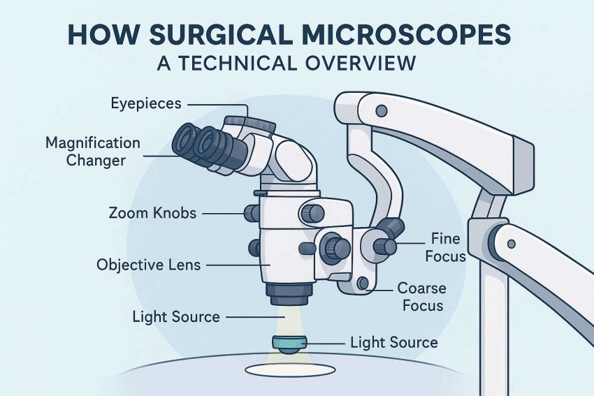
1. Objective Lens
This is the lens closest to the surgical field, responsible for collecting light reflected from the tissue.
Working Distance (WD): The space between the lens and the surgical area. Typical WDs range from 150–400 mm.
Higher-quality lenses reduce distortion and provide sharp focus even at the edges.
2. Zoom Lens System
The zoom system provides continuous magnification rather than fixed steps.
Typical range: 4x to 40x magnification.
Zoom systems can be manual or motorized (controlled by footswitch or joystick).
Zooming changes the focal length, allowing the user to enlarge or shrink the field of view without moving the microscope.
3. Eyepieces (Binocular Tubes)
Surgical microscopes use stereoscopic binocular vision, which means:
Each eyepiece receives slightly different images
The brain combines them into a 3D perception
Eyepieces often allow diopter adjustment to match the user’s vision
4. Illumination System
You can’t see what you can’t light up. Surgical microscopes use coaxial illumination to eliminate shadows and brighten deep structures.
Common light sources:
- LED (long-lasting, cool, energy-efficient)
- Xenon (brighter, whiter, used in deep cavity work)
Illumination is typically adjustable for brightness and beam focus.
Mechanical System: Movement & Positioning
Surgeons need both stability and flexibility. That’s why surgical microscopes include:
5. Articulating Arm or Stand
The stand lets the user precisely position the microscope over the surgical field.
Floor stands, ceiling mounts, or wall mounts depending on the OR layout
Counterbalanced arms allow smooth vertical and lateral movement
Locking mechanisms prevent drift during procedures
6. Foot Control
Microscopes are often controlled via foot pedals so the surgeon’s hands stay sterile.
Functions include:
- Zoom in/out
- Focus adjustment
- Light intensity control
- Switching between view modes (in digital models)
Optional Digital Features
Many high-end microscopes now include digital imaging systems for enhanced surgical precision and teaching purposes:
7. Camera & Display Integration
- Real-time video output to external monitors
- 4K or 3D image capture
- Assistants and students can follow the procedure
8. AI & Autofocus (Emerging)
- Some models include automatic focus, pattern recognition, or AR overlays
- Integration with robotic arms is becoming more common in neurosurgery and ophthalmology
Technical Specs That Matter
When evaluating a surgical microscope, key specs to understand include:
| Spec | Description |
| Magnification Range | Usually 4x–40x |
| Field of View | The width of the visible area (larger at low magnification) |
| Depth of Field | How much depth stays in focus—critical for microsurgery |
| Working Distance | Distance from lens to subject—affects ergonomics |
| Resolution | Clarity of the image—determined by optical quality |
| Illumination Intensity | Measured in lumens or lux—affects contrast and visibility |
Real-World Example: ENT Surgery
In an ENT (Ear, Nose, Throat) procedure:
- The microscope is suspended over the ear canal
- The surgeon views a 3D image through eyepieces
- The foot pedal adjusts magnification as the procedure progresses
- A coaxial LED illuminates deep inner ear structures
- A camera module records the operation for documentation or training
What’s Coming Next?
The future of surgical microscopes is moving toward:
- Digital-only viewing (exoscopes)
- Hands-free AI focus
- Augmented reality overlays
- Cloud integration for telemedicine and training
As technology evolves, we may see robot-guided optics or even surgeon-controlled interfaces through eye-tracking or voice commands.
Summary
Surgical microscopes are complex systems designed to deliver high-resolution, magnified, and well-lit views of the surgical field. They combine:
- Precision optics
- Robust mechanics
- Advanced illumination
- User ergonomics
- Optional digital technology
Understanding how they work not only empowers users—but also helps in making smarter decisions when evaluating models, comparing features, or exploring upgrades.
Lear more Surgical Microscopes Optical Components