Optical Components Explained – Understanding Lenses, Filters, and Light Paths in Surgical Microscopes
A Deep Dive Into the Core Technology That Powers Precision in Surgery
Surgical microscopes may seem like magic, but behind their precision lies the science of optics—a careful orchestration of lenses, filters, and light paths that allow surgeons to see the unseen.
In this article, we’ll explore the essential optical components of surgical microscopes, explain how each one works, and show how they contribute to image clarity, depth perception, and visual control during surgery.
Why Optics Matter in Surgery
In most surgical procedures, especially those involving delicate or deep anatomical structures, normal vision isn’t enough. Surgeons rely on microscopes to:
- Magnify small structures
- Illuminate deep cavities
- Visualize tissue layers and fine vessels
- See in stereo (3D) for depth and distance accuracy
None of this would be possible without the precise behavior of light, lenses, and optical filters.
The Core Optical Components of a Surgical Microscope
Let’s break down the main optical building blocks:
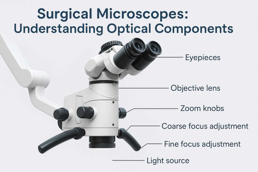
1. Objective Lens
The objective lens is the primary lens closest to the surgical field.
- It gathers light reflected from the tissue and focuses it into the optical system.
- Determines the working distance (WD), i.e., the physical distance between the lens and surgical area.
- High-quality objectives minimize chromatic aberration (color distortion) and edge blur.
Tip: Typical working distances range from 150 mm to 400 mm, depending on the type of surgery.
2. Magnification Changer (Zoom Lens System)
This is what allows adjustable magnification—either continuously (zoom) or in fixed steps (e.g., 6-step manual systems).
- Motorized zoom is standard in most high-end models.
- As magnification increases, field of view decreases and detail increases.
- The zoom system changes the focal length inside the microscope, refocusing light into a tighter or wider cone.
3. Eyepieces (Oculars)
These are where the surgeon looks through—usually a binocular tube.
- Standard magnification is 10x, with diopter adjustment to correct for vision differences.
- Stereoscopic design allows each eye to receive slightly different images, enabling depth perception (true 3D viewing).
- Interpupillary distance can be adjusted for comfort and accuracy.
Optional: Some microscopes offer tiltable binoculars or extended tubes for ergonomics in long procedures.
4. Illumination System
No optics work without light. Surgical microscopes use coaxial illumination—light that travels in the same axis as the visual path.
Types of Light Sources:
- Halogen – older tech, warm tone, limited lifespan
- Xenon – bright, white, and intense; great for deep cavity surgery
- LED – modern standard: energy-efficient, long-lasting, consistent color
Key Optical Terms:
- Light guide/fiber optic cable: channels light from the source to the objective lens
- Aperture diaphragm: controls how wide the beam is
- Field diaphragm: controls the size of the lighted area
Together, these deliver focused, shadow-free lighting exactly where the surgeon is looking.
5. Filters and Prisms
Optical Filters:
- UV Filters: block harmful ultraviolet light to protect tissue and eyes
- ND (Neutral Density) Filters: reduce brightness without altering color—useful when tissue is sensitive to light
- Green Filter: enhances visibility of red tissue contrast—popular in ENT and microsurgery
Beam Splitters and Prisms:
- Used to divide the light path for cameras, assistants, or digital displays
- A beam splitter diverts part of the image to a recording device without affecting the surgeon’s view
- Internal prisms are used to bend light and compress the microscope body for compact builds
6. Light Path Explained
Here’s a simplified diagram of the optical light path in a surgical microscope:
[Light Source] → [Fiber Cable] → [Objective Lens] → [Zoom System] → [Beam Splitter] → [Eyepieces / Camera]
The light enters through the illumination channel, reflects off the surgical site, passes through a series of optical lenses and filters, and finally reaches the eyepieces or camera.
Each element in the system has a precise alignment and coating to prevent distortion, maximize clarity, and reduce glare or reflection
| Feature | Controlled By | Result in Surgery |
|---|---|---|
| Clarity & Sharpness | Lens coatings, quality glass | Clear vision of small or deep structures |
| Depth of Field | Objective + zoom + aperture | More tissue stays in focus at once |
| Contrast | Lighting & filters | Enhanced visibility of vessels vs surrounding tissue |
| Ergonomics | Eyepiece angle, zoom range | Surgeon comfort and less fatigue |
Real-World Example: Green Filter in ENT Surgery
In ENT surgery, surgeons often activate the green filter to make red tissues (like blood vessels) stand out against surrounding structures. This enhances precision when working near critical arteries and nerves.
Similarly, in neurosurgery, neutral density filters are used to reduce glare on wet brain surfaces without lowering visibility.
Innovations in Optical Technology
Modern surgical microscopes are incorporating features like:
- Fluorescence filters – to highlight tumors or blood flow using injected dyes
- Optical Coherence Tomography (OCT) – real-time cross-sectional imaging
- Digital zoom with edge enhancement – via high-res cameras and software
- Anti-reflection coatings – reduce internal glare
Final Thoughts
Understanding the optical components of a surgical microscope helps clarify:
- Why the image quality is so detailed
- How visual contrast and focus are maintained
- Why different models are suited to different surgical applications
- Each part—from objective lens to filters—plays a vital role in turning light into precision, allowing surgeons to operate with confidence and control.